Awake craniotomy — the patient remains conscious to allow real-time mapping of brain function
Advanced TechniqueAwake Craniotomy
Brain tumour surgery performed while the patient is conscious — the gold standard for tumours near speech, language, and motor areas.
Why Awake?Maximum Safe Resection — Zero Compromise on Function
When a tumour lies near areas controlling speech or movement, conventional anaesthesia cannot guarantee function preservation. Awake craniotomy keeps the patient conscious during resection — allowing real-time functional testing. The surgeon stops the moment any boundary is approached. Result: the largest possible removal with the smallest possible neurological cost.
The ProcedureAsleep–Awake–Asleep Protocol
Three phases: the patient is anaesthetised for the opening (craniotomy), gently woken for resection while a neuropsychologist runs continuous tests (naming objects, counting, moving limbs), then returned to sleep for wound closure. Most patients find the experience far less daunting than expected — they are calm, comfortable, and actively participating in preserving their own brain function.
Cortical MappingDirect Electrical Stimulation
A tiny electrical current applied to the brain surface temporarily disrupts function in that patch of cortex — mimicking removal. Speech arrest, anomia, limb weakness, or sensory changes are detected immediately, creating a personalised functional map unique to that patient. Areas that must be preserved are clearly identified before any tissue is removed.
OutcomesResults
High-volume centres report gross total or near-total resection in 75–98% of awake craniotomy cases for glioma — significantly higher than surgery under general anaesthesia. Permanent new neurological deficit below 3–5% in experienced hands. Patients mobilise the following day and are typically discharged within 3–5 days.

Cranial neuronavigation — intraoperative GPS for the brain

Spine neuronavigation — millimetre accuracy for every case
Advanced TechnologyNeuronavigation-Guided Surgery
Intraoperative GPS for the brain and spine — millimetre precision, smaller incisions, safer outcomes every time.
Surgical GPSMillimetre Precision in Every Case
Neuronavigation registers the patient’s pre-operative MRI or CT to their actual skull position on the table. A real-time 3D display shows precisely where instruments are relative to tumour, vessels, and functional areas — allowing smaller incisions, maximum resection, and avoidance of critical structures.
Brain ApplicationsNeuronavigation in Brain Surgery
Exact tumour margins displayed in real time. The narrowest safe surgical corridor is identified before a single incision is made. Particularly valuable for deep-seated tumours, skull base lesions, recurrent tumours in distorted anatomy, and pituitary surgery guidance relative to optic chiasm and carotid arteries.
Spine ApplicationsNeuronavigation in Spine Surgery
Dramatically improves pedicle screw accuracy in complex deformity, revision, and osteoporotic spine surgery. Defines spinal tumour margins. Enables accurate MIS instrument docking through small incisions, reducing patient radiation exposure vs. repeated fluoroscopy.
CombinedNavigation + Awake Surgery + IONM
The greatest outcomes come from combining technologies. Complex tumours managed with: neuronavigation defining the target, awake craniotomy preserving function, and IONM providing continuous background safety monitoring — the current highest standard of brain tumour care.
Oncological NeurosurgeryBrain Tumour Surgery
Primary and secondary brain tumours managed with the most advanced techniques — awake craniotomy and neuronavigation as standard where indicated.
GliomaGlioma Surgery (Low & High Grade)
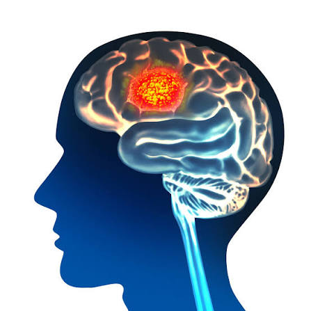 Glioma — MRI showing tumour infiltrating brain tissue
From slow-growing grade II astrocytomas to aggressive glioblastomas. Maximum safe resection guided by neuronavigation and awake cortical mapping. Extent of resection directly impacts survival — we push this to its absolute limit safely.
MeningiomaMeningioma Resection
 Meningioma — well-defined extra-axial tumour on MRI
Complete microsurgical resection (Simpson Grade I/II) offers excellent long-term control. Complex skull base meningiomas with high vascularity managed with pre-operative embolisation followed by neuronavigation-guided resection.
PituitaryPituitary Tumour — Transsphenoidal Endoscopic Surgery
 Pituitary macroadenoma on coronal MRI
Removed via the nose — no head incision required. Endoscope guided to the pituitary fossa, neuronavigation confirms safe positioning relative to optic chiasm and carotid arteries. Effective for both functioning and non-functioning adenomas.
Acoustic NeuromaAcoustic Neuroma (Vestibular Schwannoma)
 Vestibular schwannoma on MRI
Microsurgical removal targeting complete excision while preserving facial nerve function and residual hearing where possible. Intraoperative facial nerve monitoring used in every case.
MetastasisBrain Metastasis Surgery
 Multiple brain metastases on contrast-enhanced MRI
Single or limited brain metastases removed to improve survival, quality of life, and obtain tissue for molecular profiling guiding systemic oncological therapy.
Vascular NeurosurgeryCerebrovascular Surgery
Conditions affecting the brain’s blood vessels demanding rapid, expert microsurgical intervention.
AneurysmCerebral Aneurysm Clipping
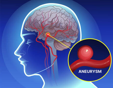
Unruptured aneurysm

Ruptured aneurysm
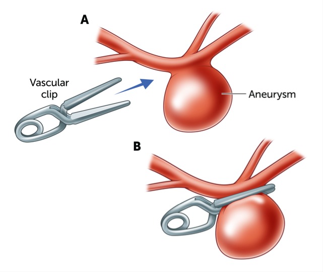
Aneurysm clipping
A metal clip placed across the aneurysm neck permanently seals it from the circulation — a definitive, one-time cure for this life-threatening condition. Both ruptured and unruptured aneurysms managed urgently.
AVMAVM Resection
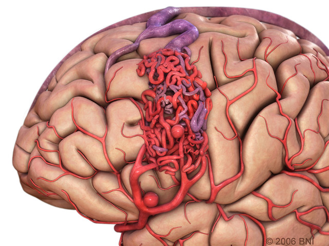 AVM — tangle of abnormal blood vessels on angiography
Microsurgical resection — sometimes combined with pre-operative embolisation — can cure the AVM entirely, eliminating future haemorrhage risk. High-grade AVMs managed with combined multimodal approach.
CavernomaCavernous Malformation Surgery
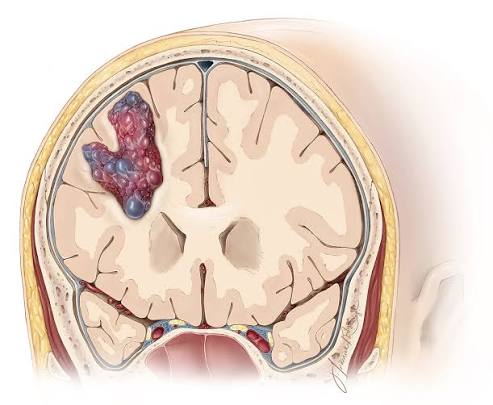 Cavernoma — popcorn-ball appearance on MRI
Symptomatic cavernomas causing repeated bleeds, progressive neurological deficits, or epilepsy are removed by microsurgery. Excellent plane allows complete removal with minimal brain disturbance.
HaemorrhageIntracerebral Haemorrhage Evacuation
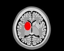 Intracerebral haemorrhage on CT scan
Large or life-threatening brain bleeds urgently evacuated via craniotomy or minimally invasive endoscopic techniques. Neuronavigation guides the most direct, least damaging route to the clot.
Emergency NeurosurgeryNeurotrauma Surgery
24/7 emergency neurosurgical availability for head injuries from minor to catastrophic. Time to surgery is the most important determinant of outcome.
TBITraumatic Brain Injury Management
 Severe TBI on CT scan
Severe TBI: rapid assessment, ICP monitoring, surgical decompression when indicated. Decompressive craniectomy for refractory raised ICP. Dr. Agarwal has extensive experience in polytrauma settings.
SubduralSubdural Haematoma Evacuation
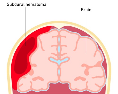 Acute subdural haematoma — crescent-shaped hyperdensity
Acute: urgent craniotomy and clot evacuation. Chronic: burr hole drainage, often under local anaesthesia. Both carry excellent outcomes when performed without delay.
ExtraduralExtradural Haematoma Surgery
 Extradural haematoma — biconvex hyperdensity on CT
A true neurosurgical emergency. Rapid craniotomy and clot evacuation — performed within the shortest possible time — is often fully curative with complete neurological recovery.
Skull FractureDepressed Skull Fracture Repair
 Depressed skull fracture on CT 3D reconstruction
Elevation and fixation of depressed fractures to prevent brain compression and infection. Cranioplasty for skull defects after decompressive craniectomy.
 Drug-resistant epilepsy — surgery can be life-changing and curative
Functional NeurosurgeryEpilepsy Surgery
For patients whose seizures are not controlled by medications — surgery can be life-changing and in many cases curative.
Temporal LobeTemporal Lobectomy / Selective Amygdalohippocampectomy
The most common and effective epilepsy surgery. Seizure freedom in 60–80% of carefully selected patients. Pre-operative video-EEG, MRI, and neuropsychological testing guide patient selection and surgical planning.
LesionectomyLesionectomy
When a visible structural lesion is the seizure source, complete removal can eliminate seizures entirely. Awake craniotomy used when the lesion abuts eloquent cortex — allowing simultaneous lesion removal and functional preservation.
Corpus CallosotomyCorpus Callosotomy
For drop-attack seizures causing dangerous falls — dividing the corpus callosum prevents seizure spread between hemispheres, dramatically reducing falls and injuries.
VNSVagus Nerve Stimulation
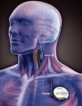 Vagus Nerve Stimulator — implanted device reduces seizure frequency
Pacemaker-like device reducing seizure frequency when resective surgery is not possible. Palliative but effective option for multifocal epilepsy.
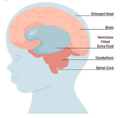 Hydrocephalus — enlarged ventricles on MRI
CSF DisordersHydrocephalus Surgery
Abnormal CSF accumulation in the brain’s ventricles — managed with surgical diversion to prevent progressive neurological damage.
VP ShuntVentriculoperitoneal (VP) Shunt
 Ventriculoperitoneal shunt — CSF drainage system
Silicone tube draining CSF from ventricles to abdomen via a programmable pressure-regulated valve. Modern valves allow non-invasive post-operative pressure adjustment without reoperation.
ETVEndoscopic Third Ventriculostomy (ETV)
 ETV — endoscopic opening at the floor of the third ventricle
A small endoscope creates an opening at the floor of the third ventricle — bypassing obstruction without an implanted device. Preferred in obstructive hydrocephalus to avoid lifelong shunt dependency.
NPHNormal Pressure Hydrocephalus
Classic triad of gait disturbance, cognitive decline, and urinary incontinence in older adults. VP shunt surgery can dramatically and often surprisingly reverse all three symptoms.
NeuromodulationDeep Brain Stimulation & Functional Surgery
Precise electrical stimulation of neurological circuits — restoring function in movement disorders, pain, and psychiatric conditions.
Parkinson’sDBS for Parkinson’s Disease
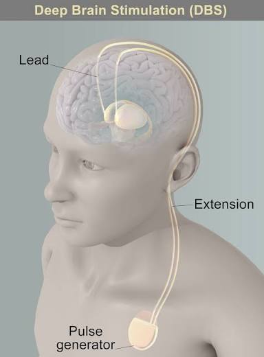 DBS electrode placement — STN or GPi targeting
Thin electrodes implanted into the STN or GPi, connected to a pulse generator. Dramatically reduces tremor, rigidity, and bradykinesia — often allowing major medication reduction. Quality of life improvement can be transformative and immediate.
Essential TremorDBS for Essential Tremor
VIM thalamic DBS — immediate tremor reduction
VIM thalamic DBS providing immediate, dramatic tremor reduction — enabling patients to eat, write, and function independently when medications have failed.
DystoniaDBS for Dystonia
GPi-DBS for generalised dystonia
GPi-DBS for generalised and focal dystonias including DYT1 genetic dystonia. Results develop gradually over weeks to months after implantation.
MVDMicrovascular Decompression (MVD)
 MVD — Teflon felt cushioning the offending vessel away from the nerve
Definitive surgical cure for trigeminal neuralgia and hemifacial spasm. The offending blood vessel is cushioned away from the nerve — immediate, durable pain relief without any nerve damage.
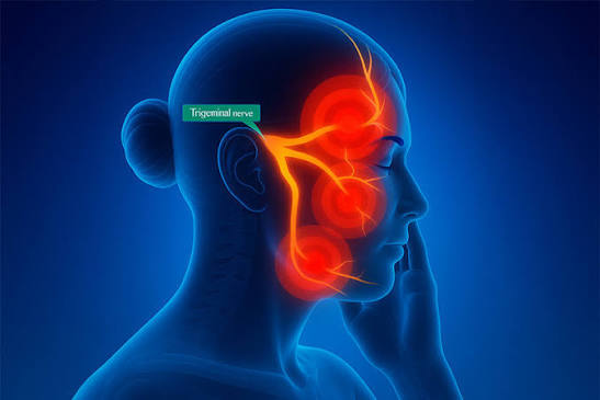 Trigeminal neuralgia — electric-shock pain along the trigeminal nerve branches
Pain SurgeryTrigeminal Neuralgia & Treatment
One of the most painful conditions known to medicine — and one of the most rewarding to treat. Dr. Agarwal has dedicated expertise in MVD surgery.
OverviewThe “Suicide Disease”
Chronic facial pain causing sudden, severe electric shock-like attacks triggered by trivial stimuli — eating, speaking, a gentle touch. Almost always caused by a blood vessel compressing the trigeminal nerve root at the brainstem. Highly treatable when the correct diagnosis is made and the right surgeon is chosen.
TypesTN1, TN2, Secondary TN
TN1: Episodic electric-shock pain with pain-free intervals — vascular compression, almost always curable with MVD. TN2: Constant background pain plus episodes — harder to treat completely. Secondary TN: MS plaques, tumours, or AVMs at the cerebellopontine angle.
DiagnosisDiagnosis
MRI with FIESTA/CISS sequences visualises neurovascular conflict and rules out secondary causes. MR Angiography maps the offending vessel. Most classical TN1 patients have a clear vascular contact on high-resolution MRI before surgery.
MedicalMedications
Carbamazepine/Oxcarbazepine first-line. Effective initially in ~70% but efficacy wanes and side effects accumulate. Medications control pain but do not address the underlying compression — surgery is curative.
Gold StandardMVD — The Definitive Cure
MVD — the definitive cure for trigeminal neuralgia
3–4cm opening behind the ear. The offending vessel — usually the superior cerebellar artery — is cushioned away from the nerve with Teflon felt. 85–90% immediate complete pain relief. 70–80% pain freedom at 10 years. No nerve damage — numbness is not an expected outcome. Discharge in 3–4 days, normal activity in 2–3 weeks.
AlternativesPercutaneous & Radiosurgical Options
Balloon microcompression, glycerol rhizotomy, radiofrequency thermocoagulation, and Gamma Knife — all available as alternatives when MVD is not feasible due to age or medical comorbidity. Each has distinct success and risk profiles discussed individually with every patient.
Peripheral Nerve SurgeryNerve Entrapment & Injury
Peripheral nerve entrapments and injuries causing significant functional disability — many surgically correctable with excellent outcomes.
Carpal TunnelCarpal Tunnel Syndrome Release
 Carpal tunnel release — decompressing the median nerve
Release of the transverse carpal ligament decompresses the median nerve. High success rate, rapid recovery, often performed as a day-case procedure.
Ulnar NerveUlnar Nerve Decompression / Transposition
 Ulnar nerve decompression at the elbow
Cubital tunnel syndrome treated by decompression or anterior transposition — freeing the nerve from its compressed position behind the medial epicondyle.
Brachial PlexusBrachial Plexus Surgery
 Brachial plexus reconstruction — nerve grafting and neurotisation
Traumatic injuries from road accidents or birth trauma treated with nerve grafting, neurotisation, or tendon transfer. Early surgical intervention achieves the best functional recovery.
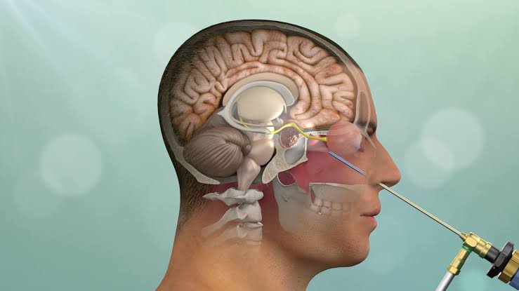 Endoscopic trans-nasal skull base repair — no external incision, no scar
Endoscopic Skull Base SurgeryEndoscopic Trans-Nasal Repair for CSF Rhinorrhoea
Cerebrospinal fluid (CSF) leaking through the nose — a potentially dangerous condition that can lead to life-threatening meningitis. Modern endoscopic repair through the nose offers definitive treatment without any external incision.
Stitchless, Scar-Free Skull Base RepairThrough the Nose — No External Incision, No Scar
CSF rhinorrhoea repair has been revolutionised by endoscopic trans-nasal techniques. Using a thin endoscope introduced through the nostril, the skull base defect is directly visualised and repaired using a layered graft technique — all without a single external incision, without shaving any hair, and without opening the skull. Patients are typically discharged within 2–3 days with a high success rate of over 90% on the first attempt.
What is CSF Rhinorrhoea?Understanding CSF Leaks
Cerebrospinal fluid (CSF) is the clear fluid that surrounds and cushions the brain and spinal cord. When there is a defect in the skull base — the bony floor separating the brain from the nasal cavity — CSF can leak out through the nose. This presents as a watery, clear nasal discharge, often described as a “dripping tap” that worsens when bending forward or straining. Why is it dangerous? The defect creates a direct communication between the brain and the nasal passages — a portal for bacteria to enter the intracranial compartment. Without repair, patients are at significant and ongoing risk of bacterial meningitis, which can be life-threatening.
CausesWhat Causes a CSF Leak?
Spontaneous (Idiopathic): The most common cause — a spontaneous defect in the skull base, often associated with raised intracranial pressure (benign intracranial hypertension/idiopathic intracranial hypertension). Patients are often overweight and present with pulsatile tinnitus and headache in addition to nasal leakage. Post-traumatic: Head injury causing a fracture through the skull base, tearing the dura. Often presents acutely after trauma but can be delayed by weeks. Post-surgical: After sinus surgery, pituitary surgery, or anterior skull base procedures. Tumour-related: Erosion of the skull base by a tumour (pituitary adenoma, meningioma, olfactory neuroblastoma) creating a defect through which CSF escapes.
DiagnosisHow is a CSF Leak Diagnosed?
Beta-2 transferrin test: The definitive biochemical test — beta-2 transferrin is found only in CSF and perilymph, not in nasal secretions. A positive test confirms CSF leak with high specificity. High-resolution CT cisternography: CT scanning after intrathecal contrast injection identifies the exact site of the skull base defect and the pathway of CSF egress. MRI with cisternography: Non-invasive; identifies active CSF leak and associated meningoceles or encephalocoeles prolapsing through the defect. Fluorescein intrathecal injection: Used intraoperatively — fluorescein dye injected into the spinal fluid glows bright yellow-green under blue light, making even the smallest defect immediately identifiable during endoscopic repair.
The Endoscopic ProcedureEndoscopic Trans-Nasal Skull Base Repair — Step by Step
Under general anaesthesia, a rigid endoscope is introduced through the nostril. The nasal cavity is carefully examined and the site of leakage is identified — often aided by intrathecal fluorescein glowing at the defect site. The mucosa around the defect is carefully removed to create a fresh edge for graft adherence. A layered repair is then constructed: Layer 1 — Underlay: A piece of fat, fascia lata (thigh fascia), or nasal turbinate mucosa is placed intracranially through the defect to plug it from the inside. Layer 2 — Overlay: A second graft (nasal septal flap, middle turbinate mucosa, or synthetic dural substitute) is placed over the defect on the nasal side and secured with fibrin glue. A nasal pack is placed for 3–5 days to support the repair. Success rates exceed 90% with a single procedure; most residual cases are successfully treated with a second endoscopic attempt.
Special CasesMeningoceles & Encephalocoeles
In some patients — particularly those with longstanding elevated intracranial pressure — the brain lining (meninges) or even brain tissue itself herniates through the skull base defect, creating a meningocele or encephalocoele. These require careful endoscopic reduction of the herniated contents back into the skull, followed by a multi-layer repair of the defect. Simultaneous management of the underlying raised intracranial pressure (with medication or CSF diversion) is essential to prevent recurrence.
RecoveryWhat to Expect
Hospital stay: typically 3–5 days. Nasal packing removed at 3–5 days. Patients are asked to avoid nose-blowing, heavy lifting, straining, and any Valsalva manoeuvre for 4–6 weeks to allow the repair to heal. No external stitches, no visible scar. Success rate >90% after first repair. Return to normal activity: 3–4 weeks. Associated idiopathic intracranial hypertension is managed concurrently with acetazolamide and weight loss programme to reduce the risk of recurrence.
Reconstructive & Aesthetic NeurosurgeryCranioplasty & Skull Surgery
Skull defects and deformities — from trauma, previous surgery, or congenital conditions — cause both functional concerns and significant cosmetic distress. We offer the full spectrum of cranial reconstruction and beautification.
Aesthetic & Functional Skull ReconstructionRestoring the Skull — Restoring Confidence
A skull defect or deformity is not merely a cosmetic concern. It can leave the brain unprotected, cause headaches and neurological symptoms, and profoundly affect a patient’s self-image and quality of life. At our centre, we specialise in the full spectrum of cranial reconstruction — from emergency cranioplasty after decompressive surgery to elective correction of congenital and acquired skull deformities. Every patient receives a customised surgical plan designed for the best possible functional and aesthetic outcome.
CranioplastyCranioplasty — Skull Defect Repair
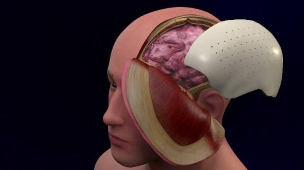 Cranioplasty — custom PEEK implant reconstruction
After decompressive craniectomy (emergency surgery for severe brain swelling), a significant skull defect remains. Cranioplasty repairs this defect, protecting the brain and restoring normal intracranial pressure dynamics. Materials used: Autologous bone (the patient’s own skull bone, stored after decompression), titanium mesh, or custom-made PEEK (polyether ether ketone) implants designed from CT scans for a perfect anatomical fit. Custom PEEK implants provide the most aesthetically superior result and are used in complex cases requiring precise contour matching.
Fibrous DysplasiaFibrous Dysplasia — Skull Contour Correction
 Fibrous dysplasia — abnormal bone expansion with skull deformity
What is fibrous dysplasia? A bone disorder where normal bone is replaced by fibrous tissue, causing progressive bone expansion, deformity, and abnormal contour. In the skull, this creates visible lumps, asymmetry, orbital deformity, and sometimes neurological compromise from narrowed nerve exit channels (foramina). Surgery: Affected bone is carefully resected and the skull contour is reconstructed using custom titanium or PEEK implants, hydroxyapatite cement, or a combination. The result is a smooth, natural skull contour with functional and aesthetic restoration. Orbital involvement causing visual loss or proptosis is managed in collaboration with ophthalmology and orbital surgery teams.
Skull Base ReconstructionSkull Base Reconstruction After Tumour Surgery
After skull base tumour surgery, significant reconstruction may be needed to seal the dura, obliterate dead space, and restore the skull base architecture. We use pericranial flaps, fat grafts, titanium mesh, and PEEK implants — preventing CSF leak, meningitis, and cranial nerve dysfunction after extensive skull base resections.
Aesthetic ContouringCosmetic Skull Contouring
Asymmetries, indentations, and abnormal prominences of the skull — whether congenital or acquired — can be corrected with bone grafting, hydroxyapatite cement, or custom implants. We work with each patient to define their aesthetic goals and achieve a natural, symmetric result through the smallest possible incision.
Post-TraumaPost-Traumatic Skull Reconstruction
Road traffic accidents, assaults, and falls can cause skull fractures with loss of bone, cosmetic deformity, and risk of injury to the underlying brain. Delayed skull reconstruction using custom implants restores both protection and appearance, often dramatically improving a patient’s self-confidence and quality of life.
Advanced NeurosurgerySkull Base Surgery
The skull base is the floor of the cranial cavity — a complex anatomical region where the brain interfaces with the face, orbits, sinuses, and neck. Tumours and conditions here are among the most technically challenging in all of surgery. Our centre offers the full spectrum of skull base approaches, combining neuronavigation, endoscopy, and microsurgery for optimal outcomes.
One of the Most Complex Disciplines in NeurosurgeryWhere Anatomy, Technology & Surgical Skill Converge
Skull base surgery requires mastery of intricate anatomy involving the optic nerves, carotid arteries, cranial nerves, brainstem, and venous sinuses — all packed into a confined space. At our centre, we use neuronavigation, intraoperative neurophysiological monitoring, endoscopy, and microsurgical technique in combination to achieve safe, complete resection of skull base pathology. When the world declares a skull base tumour inoperable — we plan differently.
Anterior Skull BaseAnterior Skull Base Tumours
 Anterior skull base tumour on MRI
Olfactory groove meningiomas: Large meningiomas growing along the floor of the anterior cranial fossa, often presenting with personality change, visual loss, and anosmia. Microsurgical resection through a bifrontal or supraorbital approach achieves complete removal in most cases. Planum sphenoidale meningiomas: Arising on the flat bone behind the cribriform plate, often involving the optic canals. Early surgery before irreversible visual loss is critical. Olfactory neuroblastoma (Esthesioneuroblastoma): Malignant tumours arising in the olfactory epithelium, extending through the cribriform plate into the anterior cranial fossa. Managed with combined craniofacial resection — often endoscopically assisted — and adjuvant therapy.
Middle Skull BaseMiddle Skull Base & Cavernous Sinus
 Middle skull base — cavernous sinus region tumour
Cavernous sinus meningiomas: One of the most challenging skull base locations, involving cranial nerves III, IV, V, and VI alongside the carotid artery. Management is individualised — surgery, radiosurgery, or combined — with the goal of preserving cranial nerve function. Trigeminal schwannomas: Benign tumours of the trigeminal nerve often extending through the skull base. Complete microsurgical removal via a middle fossa or combined approach. Pituitary macroadenomas with suprasellar/cavernous extension: Large adenomas extending beyond the sella into the skull base require endoscopic transsphenoidal plus, in selected cases, transcranial resection.
Posterior Skull Base / CPAPosterior Skull Base & Cerebellopontine Angle (CPA)
CPA tumour — acoustic neuroma on MRI
The cerebellopontine angle is the most common site of posterior skull base tumours. Acoustic neuromas (vestibular schwannomas): Arising on the vestibular nerve, managed via retrosigmoid, translabyrinthine, or middle fossa approaches depending on tumour size and hearing status. Intraoperative facial nerve monitoring is mandatory. CPA meningiomas: Meningiomas in this location are resected via retrosigmoid craniotomy, with cranial nerve monitoring throughout. Epidermoid cysts, arachnoid cysts: Benign lesions causing cranial nerve symptoms; excellent results with microsurgical removal. Glomus jugulare tumours: Highly vascular tumours of the jugular foramen managed with pre-operative embolisation followed by microsurgical resection.
Clival & Foramen MagnumClival Tumours & Foramen Magnum
 Clival chordoma on MRI sagittal view
Chordomas: Locally aggressive tumours arising from notochordal remnants in the clivus. Complete surgical resection is the only chance of cure — achieved via endoscopic transnasal, transcervical, or combined craniofacial approaches. Chondrosarcomas: Cartilaginous tumours at the skull base requiring aggressive surgical resection. Foramen magnum meningiomas: Meningiomas at the junction of the skull and cervical spine causing brainstem compression. Resected via a far-lateral posterior fossa craniotomy — one of the most demanding approaches in skull base surgery. Craniovertebral junction instability: Managed with posterior occipitocervical fusion when the skull base or upper cervical spine is rendered unstable by tumour, trauma, or rheumatoid disease.
Endoscopic Skull BaseEndoscopic Skull Base Surgery
Many skull base tumours previously requiring large open craniotomies can now be addressed entirely through the nose using extended endoscopic transsphenoidal approaches. Conditions amenable to purely endoscopic skull base resection: Pituitary adenomas, craniopharyngiomas, clival chordomas, selected anterior skull base meningiomas, CSF leaks, and selected olfactory neuroblastomas. The endoscope provides panoramic illumination of the skull base, allowing safe work around critical neurovascular structures without brain retraction. Neuronavigation is used in every endoscopic skull base case.
Approach SelectionChoosing the Right Approach
Skull base surgery is not a single operation — it is a discipline with dozens of distinct surgical approaches, each designed to provide optimal access to a specific anatomical region while minimising brain and cranial nerve disturbance. The choice of approach depends on tumour location, size, vascularity, involvement of cranial nerves and vascular structures, and the patient’s hearing status and functional priorities. At our centre, every skull base case is discussed in a multidisciplinary team meeting before surgery to determine the optimal strategy. We believe that careful pre-operative planning is as important as the surgery itself.
Paediatric NeurosurgeryPaediatric Neurosurgery
Children are not small adults — their neurological conditions, surgical anatomy, and recovery physiology are unique. Dr. Agarwal has dedicated experience in paediatric neurosurgery, managing a wide spectrum of conditions from birth defects to brain tumours in the youngest patients.
Special Expertise & Compassionate CareEvery Child Deserves the Best Start
Paediatric neurosurgical conditions require not only technical mastery but also deep sensitivity to the family's anxiety and the child's unique needs. Our team works closely with paediatric anaesthesiologists, neonatologists, and paediatric intensivists to provide comprehensive, family-centred neurosurgical care. We understand that operating on a child's brain or spine is one of the most significant events a family can face — and we are committed to making the journey as safe, clear, and supported as possible.
Brain TumoursPaediatric Brain Tumours
Paediatric brain tumour on MRI
Medulloblastoma: The most common malignant paediatric brain tumour, arising in the cerebellum. Microsurgical resection followed by craniospinal radiotherapy and chemotherapy. Maximum safe resection significantly improves survival. Ependymoma: Arises from the lining of the ventricular system, commonly in the posterior fossa. Complete surgical resection is the most important prognostic factor. Pilocytic astrocytoma: The most common paediatric brain tumour overall — typically benign and often completely curable with surgical resection alone. Craniopharyngioma: A benign but challenging tumour near the pituitary and optic chiasm causing hormonal, visual, and cognitive problems. Management requires careful balance between extent of resection and preservation of hypothalamic function. DIPG (Diffuse Intrinsic Pontine Glioma): A devastating brainstem tumour currently managed with radiotherapy; surgical biopsy for molecular profiling is now recommended to guide targeted therapy.
HydrocephalusPaediatric Hydrocephalus
 Paediatric hydrocephalus — enlarged head circumference
Hydrocephalus is more common in children than adults, with unique causes including aqueductal stenosis, post-haemorrhagic hydrocephalus of prematurity, Chiari malformation, tumours, and infection. Management with VP shunts uses paediatric-sized catheters, programmable valves, and growth-accommodating designs. Endoscopic Third Ventriculostomy (ETV) is particularly effective in older children with obstructive hydrocephalus, avoiding lifelong shunt dependency.
Epilepsy SurgeryPaediatric Epilepsy Surgery
Drug-resistant childhood epilepsy — early surgery preserves development
Drug-resistant epilepsy in children is particularly devastating — impacting cognitive development, schooling, and quality of life with every uncontrolled seizure. Early surgical evaluation is critical. Paediatric epilepsy surgery includes temporal lobectomy, hemispherectomy (for hemispheric disorders), corpus callosotomy, and lesionectomy. The earlier a surgical cure is achieved, the better the chance of preserving normal developmental trajectory.
CraniosynostosisCraniosynostosis
 Craniosynostosis — premature fusion of skull sutures
Premature fusion of the skull sutures restricts brain growth and causes characteristic skull deformities (scaphocephaly, trigonocephaly, plagiocephaly). Surgical correction in infancy — either open cranial vault remodelling or endoscope-assisted strip craniectomy — releases the restriction and allows normal brain development. Early surgery, ideally before 6 months of age, gives the best cosmetic and neurological outcomes.
Spinal CordPaediatric Spinal Cord Conditions
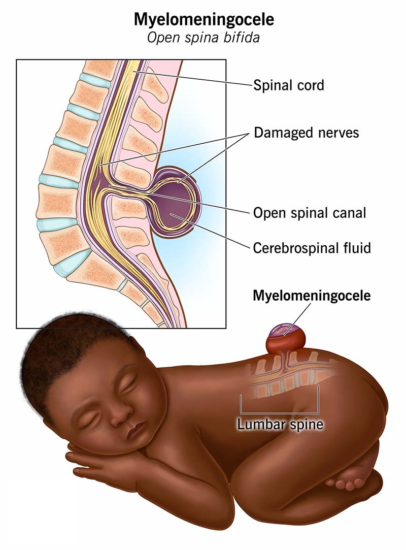 Tethered cord and spinal dysraphism in children
Tethered spinal cord, spinal cord tumours (ependymomas, astrocytomas), diastematomyelia (split spinal cord malformation), and lipomyelomeningocele are managed with careful microsurgical technique under IONM. The goal is always to preserve or improve neurological function while preventing further deterioration as the child grows.
Congenital NeurosurgeryNeural Tube Defects & Congenital Brain Conditions
Neural tube defects result from failure of the neural tube to close during early foetal development. These conditions range from benign to life-threatening and require skilled neurosurgical management — often in the first hours to days of life.
From Birth — Expert Care for Complex Congenital ConditionsEarly Surgery, Lifelong Impact
Congenital brain and neural tube conditions require neurosurgeons experienced in neonatal anatomy, rapid stabilisation, and family counselling. Our centre manages the full spectrum — from emergency neonatal repair of open neural tube defects to elective surgery for Chiari malformations and encephalocoeles — in close collaboration with neonatology, paediatrics, and rehabilitation teams.
EncephalocoeleEncephalocoele
 Occipital encephalocoele — herniation of brain tissue through skull defect
An encephalocoele is a herniation of brain tissue and meninges through a defect in the skull. It may be occipital (most common in Asia), frontonasal (sincipital), or basal. Occipital encephalocoele: Presents as a sac at the back of the head, often containing dysplastic brain tissue. Surgical repair involves excision of the sac, reduction of herniated contents where possible, and watertight closure of the dura and scalp. Frontonasal (sincipital) encephalocoele: Presents as a mass between the eyes or at the root of the nose. Repair requires a combined intracranial and extracranial approach to achieve complete removal and reconstruct the skull base. The cosmetic result is closely attended to. Basal encephalocoele: Herniates into the nasal cavity or nasopharynx, often undiagnosed until later childhood. Endoscopic transnasal repair is highly effective.
Chiari MalformationChiari Malformation
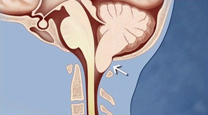 Chiari malformation — tonsillar herniation on sagittal MRI
In Chiari malformation, the cerebellar tonsils herniate downward through the foramen magnum into the spinal canal, causing headache (especially on coughing/straining), neck pain, dizziness, swallowing difficulties, arm/hand numbness, and progressive myelopathy. Chiari I: Tonsillar herniation >5mm without spinal cord or brain structural abnormality. Surgery (posterior fossa decompression — suboccipital craniectomy and C1 laminectomy) is indicated when symptomatic. The dura is opened and a dural patch inserted to expand the posterior fossa. Associated syringomyelia (fluid cavity in the spinal cord) usually resolves after decompression. Chiari II: Associated with myelomeningocele, nearly always symptomatic from birth. Requires urgent evaluation and often surgical treatment. Chiari III: Rare and severe; involves herniation of the cerebellum and brainstem into a large encephalocoele.
Dandy-WalkerDandy-Walker Malformation
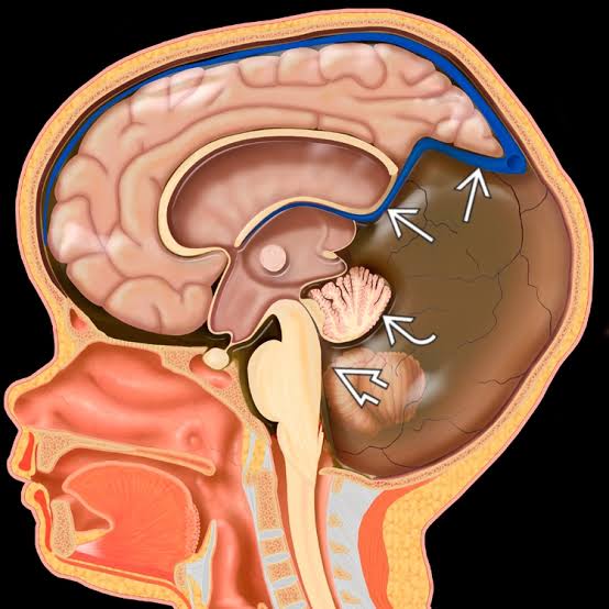 Dandy-Walker malformation — large posterior fossa cyst on MRI
A spectrum of posterior fossa malformations involving cystic enlargement of the fourth ventricle, partial or complete absence of the cerebellar vermis, and an enlarged posterior fossa. Commonly associated with hydrocephalus. Management involves CSF diversion (VP shunt or cystoperitoneal shunt) for hydrocephalus, and multidisciplinary developmental support.
Arachnoid CystsIntracranial Arachnoid Cysts
 Middle fossa arachnoid cyst on MRI
Collections of CSF within the arachnoid membrane, most commonly in the middle cranial fossa (temporal), suprasellar, posterior fossa, or convexity. Many are asymptomatic and require only observation. Symptomatic cysts causing headache, seizures, developmental delay, or hydrocephalus are treated by endoscopic fenestration (creating a window into the cyst to allow CSF reabsorption) or cystoperitoneal shunting.
Corpus CallosumAgenesis of the Corpus Callosum
 Complete agenesis of corpus callosum on MRI
Partial or complete failure of development of the corpus callosum — the main white matter tract connecting the two hemispheres. Often discovered incidentally or presenting with seizures, developmental delay, or cognitive differences. Surgery is not indicated for the callosal agenesis itself but may be required for associated hydrocephalus, seizures, or other structural abnormalities.
 Minimal to no hair shaving — preserving identity throughout the surgical journey
Patient-Centred CareHair-Sparing Brain Surgery
We understand that hair is an integral part of a patient's appearance, identity, and self-confidence. At our centre, we are committed to preserving as much hair as possible for every cranial surgery — because looking and feeling like yourself matters, even during the most difficult medical journey.
Our PromiseMinimal to No Hair Shaving — For Every Cranial Surgery
Gone are the days of shaving an entire head for brain surgery. With modern surgical planning, precise incision design, and careful hair management techniques, we routinely perform craniotomies with only a narrow strip of hair removed along the planned incision line — and in many cases, virtually no shaving at all. The surrounding hair is parted, kept intact, and can be styled to conceal the incision site almost immediately after surgery.
We believe that hair is an integral part of a patient's look and personality — and we respect and preserve it to the maximum extent that surgical safety allows. Patients have told us that this seemingly small consideration made an enormous difference to how they experienced their recovery.
How We Do ItThe Hair-Sparing Technique
Pre-operative planning: The incision is planned using neuronavigation to identify the shortest, most direct route to the surgical target — minimising incision length and therefore the amount of hair affected. Minimal strip shaving: Only a narrow 1–2cm strip directly along the planned incision line is shaved. All surrounding hair is left intact. Parting and securing: Hair on either side of the incision is carefully parted and secured away from the operative field using clips and drapes — keeping it clean and tangle-free throughout the procedure. Post-operative styling: Because only a narrow strip is shaved, the incision can be concealed within days by the surrounding hair for most patients — particularly those with medium to long hair. Patient feedback: Patients consistently report that minimal hair shaving significantly reduced their anxiety before surgery and helped them feel more like themselves during recovery.
Which ProceduresApplicable to Almost All Cranial Surgeries
Hair-sparing technique is applicable to the vast majority of elective cranial surgeries including brain tumour surgery, awake craniotomy, AVM resection, aneurysm clipping, epilepsy surgery, DBS implantation, skull base surgery, cranioplasty, and decompressive craniectomy. In true emergencies where speed is paramount, safety always takes precedence — but even in urgent cases we aim to minimise unnecessary hair removal.
For All PatientsThis Matters for Every Age & Gender
We are especially mindful of this for women, young patients, and anyone for whom hair is a particularly important aspect of identity. But we apply the same hair-sparing philosophy universally — because dignity and self-image matter for every patient, regardless of age or gender. We also counsel patients pre-operatively about what to expect, so there are no surprises when they look in the mirror after surgery.
Neuro-InfectionsBrain Infections & Tuberculosis
India carries one of the world’s highest burdens of central nervous system tuberculosis. From pyogenic abscesses to intracranial tuberculomas, timely diagnosis and the right balance of medical and surgical treatment are critical to preserving neurological function.
Medical + SurgicalMost CNS Infections Are Cured — When Treated Early
The majority of brain infections respond to a carefully chosen combination of targeted antimicrobial or anti-tubercular therapy and, where indicated, minimally invasive neurosurgical drainage. The neurosurgeon’s role is to relieve dangerous pressure, obtain tissue for accurate diagnosis (bacterial vs. tubercular vs. fungal), and treat complications such as hydrocephalus — while the infection itself is eradicated with medication. Speed matters: outcomes are excellent when treatment begins before irreversible neurological damage occurs.
Brain AbscessPyogenic Brain Abscess
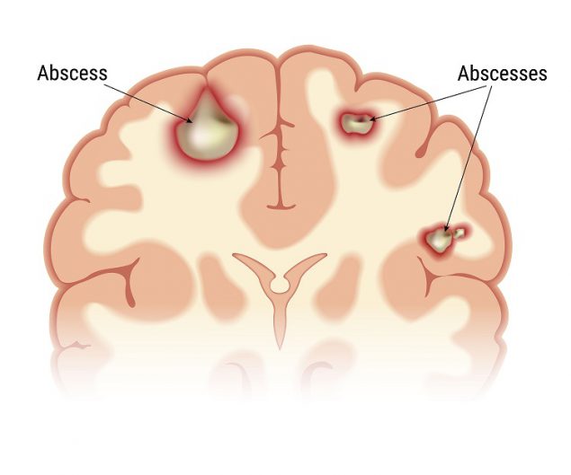 Brain abscess — ring-enhancing collection of pus
A localised collection of pus within the brain, often spreading from ear/sinus infection, dental sepsis, or following trauma or surgery. Presents with headache, fever, seizures, or focal deficits. Treatment: neuronavigation- or stereotactic-guided aspiration through a small burr hole drains the pus, confirms the organism, and relieves pressure — followed by a prolonged course of targeted antibiotics. Larger or multiloculated abscesses may require craniotomy and excision.
EmpyemaSubdural & Extradural Empyema
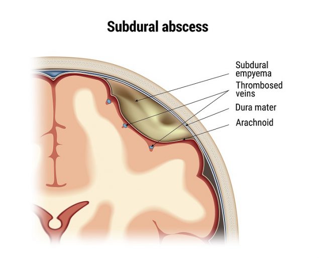 Subdural empyema — pus between the brain coverings
A neurosurgical emergency — pus collecting between the brain coverings, usually from sinusitis or middle-ear infection. Rapid neurological deterioration is common. Treatment: urgent surgical evacuation via burr holes or craniotomy to drain the collection and decompress the brain, combined with high-dose intravenous antibiotics and treatment of the underlying source. Early surgery is the single most important factor for full recovery.
TuberculomaIntracranial Tuberculoma
 Intracranial tuberculoma on MRI
A tuberculous granuloma within the brain that can mimic a tumour on imaging. Treatment is primarily medical — a full course of anti-tubercular therapy (ATT), often producing complete resolution. Neurosurgery is reserved for diagnostic biopsy when the nature is uncertain, or for large lesions causing significant mass effect, raised pressure, or seizures unresponsive to medication.
TB MeningitisTubercular Meningitis & Hydrocephalus
TB meningitis causing hydrocephalusTubercular meningitis frequently obstructs the normal flow of cerebrospinal fluid, causing hydrocephalus and dangerously raised intracranial pressure. Treatment: alongside anti-tubercular therapy and steroids, the pressure is relieved surgically with a VP shunt or an endoscopic third ventriculostomy (ETV). Prompt CSF diversion can be life-saving and dramatically improves the level of consciousness and long-term neurological outcome.
Fungal & OtherFungal Granuloma & Opportunistic Infections
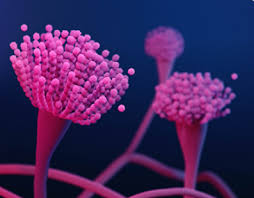 Fungal infection involving the brain and skull base
In diabetics and immunocompromised patients, fungal infections (such as aspergilloma or mucormycosis extending from the sinuses) can involve the skull base and brain. Management combines aggressive surgical debridement, antifungal therapy, and control of the underlying condition. A multidisciplinary approach with ENT, infectious-disease, and critical-care teams gives the best chance of survival and recovery.