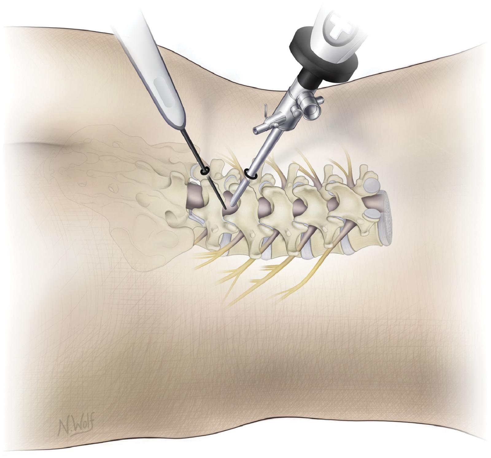
Biportal endoscopy — dual portal technique
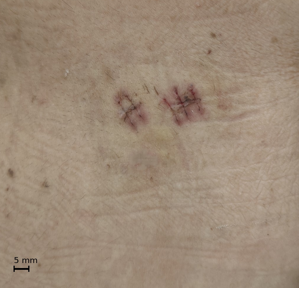
Extremely small 0.5 cm portals — minimal scarring
Ultra Minimally Invasive · SpecialisationBiportal Endoscopic Spine (UBE)
Unilateral Biportal Endoscopy (UBE) is the most advanced minimally invasive spine surgery available. Dr. Agarwal is among the select spine surgeons in Delhi NCR with dedicated training in this technique.
Two Portals. Full Freedom. Minimal Trauma.The Next Generation of Minimally Invasive Spine Surgery
Unlike uniportal endoscopy where camera and instruments share one channel, UBE uses two independent 1cm portals — one for a high-definition arthroscope, one for instruments. Complete freedom of instrument movement independent of the camera, replicating open microsurgery dexterity through two tiny skin punctures. Continuous saline irrigation provides outstanding, blood-free visualisation throughout. Most patients admitted day of surgery and home in 24–48 hours.
The TechniqueHow Biportal Endoscopy Works
4mm arthroscope delivers wide-angle HD vision through one portal. Instruments enter independently through the second — free triangulation onto any structure. Saline irrigation automatically clears blood. Instruments pass between muscle fibres without cutting — post-operative enzyme levels dramatically lower than even tubular MIS surgery. Far less pain, faster recovery.
ProceduresWhat Can Be Done Through Two Tiny Portals?
UBE Discectomy: Herniated disc removed with open-surgery thoroughness through two 1cm incisions. ULBD: Entire spinal canal decompressed through one side for stenosis. BE-TLIF: Full interbody fusion including cage and pedicle screws through two portals. Cervical foraminotomy: Motion-preserving decompression without fusion. Intradural pathology: Selected tumours accessible endoscopically.
AdvantagesUBE vs. All Other Approaches
vs. Open: Far less muscle damage, blood loss, pain — faster return to work. vs. Uniportal endoscopy: Greater surgical freedom, wider range including fusion, better for complex cases. vs. Tubular MIS: Superior illumination, less retraction, automatic blood clearance — cleaner field throughout.
RecoveryDischarge & Recovery Timeline
Discectomy and decompression: walk within hours, discharged 24–48h, desk work in 1–2 weeks. BE-TLIF fusion: walking within 24h, discharged 2–3 days, back to normal in 3–4 weeks. Physical work: 4–6 weeks for all procedures.
 Full-endoscopic uniportal spine surgery — single-incision, walk-out recovery
Fellowship TrainedUniportal Endoscopic Spine Surgery
Full-endoscopic disc surgery — the least invasive single-incision option. Walk-out-the-door recovery for most patients.
TESSYSFull-Endoscopic Transforaminal Lumbar Discectomy
A 7–8mm working channel endoscope through a small flank skin puncture. No muscle stripping, no bone removal, often under local anaesthesia. Walk the same day, home within 24 hours.
CESSYSFull-Endoscopic Cervical Discectomy
Posterior cervical foraminotomy and discectomy for arm pain from cervical disc. Avoids fusion entirely — full preservation of neck motion through a single small incision.
InterlaminarFull-Endoscopic Interlaminar Approach
Alternative route for central and paracentral disc herniations at L4-L5 and L5-S1. Excellent visualisation with minimal tissue trauma.
StenosisEndoscopic Decompression for Lumbar Stenosis
Spinal stenosis causing neurogenic claudication treated endoscopically — no destabilisation, no fusion. Walk-out recovery in suitable patients.
Minimally InvasiveMIS Spine Surgery
Tubular and percutaneous techniques minimise muscle damage, reduce blood loss, and shorten recovery.
MIS-TLIFMinimally Invasive TLIF
 MIS-TLIF — percutaneous pedicle screw fixation
For spondylolisthesis, degenerative disc disease, recurrent disc herniation requiring fusion. Two small incisions replace a long midline opening — less pain, faster return to activity.
TubularTubular Microdiscectomy
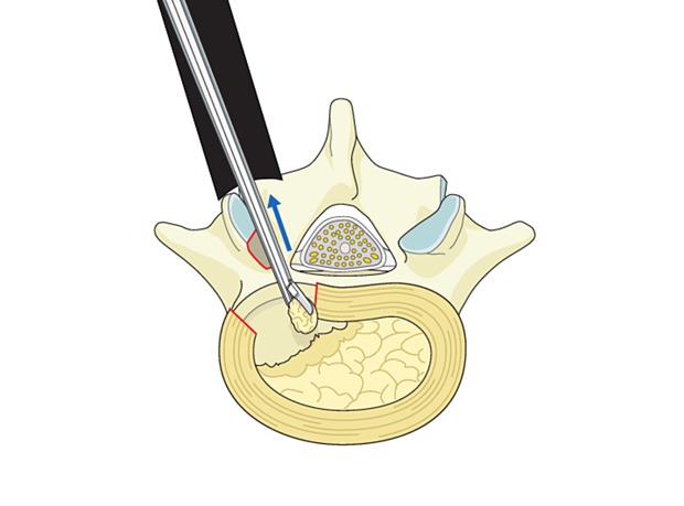 Tubular microdiscectomy — 18mm retractor, microscopic precision
18mm tubular retractor under microscopic vision. Less muscle damage than open discectomy with equivalent or superior outcomes.
PercutaneousPercutaneous Pedicle Screw Fixation
Percutaneous pedicle screw fixation — stab incisions only
Spinal fusion hardware through tiny stab incisions under navigation or fluoroscopic guidance. No large muscle stripping of open instrumentation.
VertebroplastyVertebroplasty / Kyphoplasty
 Vertebroplasty — fluoroscopic-guided cement injection
Stitchless, needle-based procedure for painful vertebral compression fractures. Immediate pain relief. Same-day or next-day discharge. See full details in the Vertebroplasty / Kyphoplasty tab above.
Cervical SpineCervical Spine Surgery
From motion-preserving endoscopic decompression to multi-level reconstruction — tailored to each patient’s anatomy and goals.
ACDFAnterior Cervical Discectomy & Fusion (ACDF)
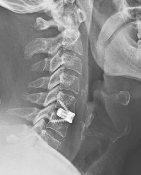 ACDF — cage and plate fixation
Standard operation for cervical disc herniation and spondylosis causing radiculopathy or myelopathy. Small neck incision, disc replaced with cage and plate. Typically discharged within 1–2 days.
CDRCervical Disc Replacement
 Cervical disc replacement — motion-preserving arthroplasty
Artificial disc preserving cervical motion and reducing adjacent segment disease risk. Ideal for younger patients with single-level disc disease and good bone quality.
LaminoplastyCervical Laminoplasty
 Cervical laminoplasty — canal expansion with open-door technique
For multilevel cervical stenosis causing myelopathy — canal expansion by hinging laminae open. Motion-preserving alternative to laminectomy and fusion.
PosteriorPosterior Cervical Fusion
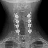 Posterior cervical fusion — lateral mass screw fixation
For multilevel instability or failed anterior surgery — posterior fixation with lateral mass or pedicle screws provides definitive multi-level stability.
Lumbar SpineLumbar Spine Surgery
The most common source of disability worldwide — treated with a precise, individualised approach from endoscopic to open.
MicrodiscectomyLumbar Microdiscectomy
Lumbar microdiscectomy — microscopic precision
Gold standard for disc herniation causing sciatica. Microscopic precision, minimal collateral damage. Most patients home within 24–48h and walking comfortably.
LaminectomyLumbar Laminectomy / Decompression
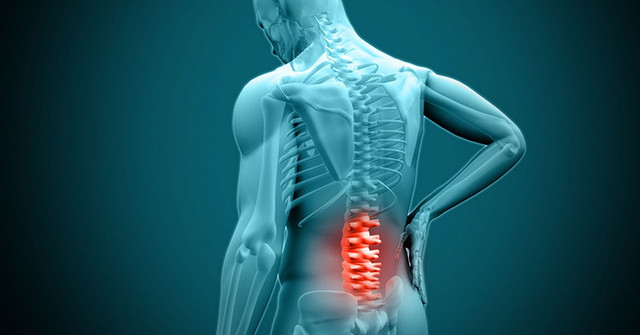 Lumbar decompression — relieving spinal canal stenosis
Removal of lamina and hypertrophied ligament to decompress the spinal canal in stenosis. Reliable, durable relief of neurogenic claudication (leg pain on walking).
TLIF/PLIFTLIF / PLIF (Interbody Fusion)
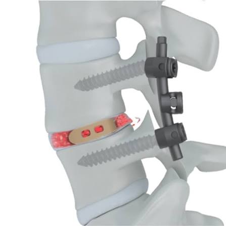 TLIF/PLIF — interbody cage with pedicle screw fixation
For instability, spondylolisthesis, or recurrent disc disease requiring stabilisation. Interbody cage plus pedicle screw fixation. Preferred as MIS-TLIF where possible.
ALIFALIF (Anterior Lumbar Interbody Fusion)
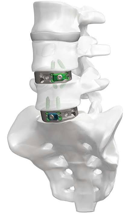 ALIF — anterior approach for L5-S1 fusion
Fusion through an abdominal approach — larger graft, better disc height restoration, particularly effective at L5-S1 where posterior access is limited.
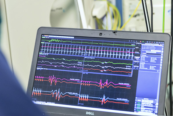 Intraoperative Neurophysiological Monitoring (IONM) — standard of care for all spinal tumour surgery
Spinal OncologySpinal Tumour Surgery
All spinal tumour surgeries at our centre performed with Intraoperative Neurophysiological Monitoring (IONM) as the standard of care — the most important safety advancement in spinal tumour surgery.
Our Standard of Care — IONMReal-Time Neural Monitoring Throughout Every Spinal Tumour Surgery
A dedicated neurophysiologist continuously records electrical signals via scalp, limb, and muscle electrodes throughout the operation. Three modalities run simultaneously:
SSEPs: Any cord compression causes immediate waveform changes — detected before a permanent sensory deficit develops.
MEPs: Monitor motor pathway (corticospinal tract) integrity in real time — detect motor compromise before movement is affected.
EMG: Free-running and triggered EMG monitors individual nerve root function during every moment of retraction and instrumentation.
When a signal change is detected, the surgeon immediately modifies the approach — releasing retraction, correcting blood pressure, adjusting technique — often reversing the change completely before any permanent damage. Mandatory for intramedullary cord tumours at all high-volume centres. Standard of care for every spinal tumour case at our centre.
IDEMIntradural Extramedullary Tumours — Meningioma & Schwannoma
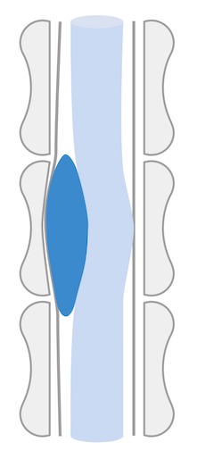 IDEM tumour — meningioma/schwannoma on spinal MRI
Benign tumours outside the cord but inside the dura. Microsurgical removal under continuous IONM — highly effective, often curative, with excellent neurological recovery. The plane between tumour and cord allows complete removal in most cases.
IntramedullaryIntramedullary Spinal Cord Tumours
 Intramedullary tumour — ependymoma within the spinal cord
Ependymomas and astrocytomas within the spinal cord — the most technically demanding spinal surgery. Midline myelotomy, tumour removed under continuous MEP, SSEP, and EMG guidance. Every moment of resection guided by IONM feedback. Surgeon stops or modifies immediately if signals change. This approach maximises tumour removal while minimising permanent deficits.
ExtraduralExtradural Spinal Metastases
 Extradural metastasis causing cord compression
Cord compression from metastatic disease requiring urgent decompression and stabilisation. IONM guides decompression and confirms adequacy in real time. Surgery restores neurological function and allows adjuvant treatment to proceed.
Spinal EmergencySpinal Trauma Surgery
Traumatic spinal injuries demanding urgent assessment, immobilisation, and surgical stabilisation to maximise neurological recovery.
Fracture FixationVertebral Fracture Fixation
 Vertebral fracture — pedicle screw fixation
Unstable thoracolumbar and cervical fractures: pedicle screw instrumentation and fusion to restore alignment, decompress cord, and allow early mobilisation.
SCISpinal Cord Injury Management
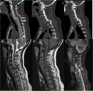 Spinal cord injury — urgent decompression and stabilisation
Emergency decompression within hours of incomplete SCI can meaningfully improve neurological outcomes. Dr. Agarwal has extensive experience managing acute SCI and coordinating multidisciplinary rehabilitation.
Upper CervicalOdontoid Fracture / Atlanto-axial Stabilisation
 Atlanto-axial instability — C1-C2 fracture on CT
C1-C2 fractures — particularly dangerous. Posterior atlanto-axial fusion with Harms technique or anterior odontoid screw fixation provides definitive stability preventing catastrophic cord injury.
Stitchless Minimally InvasiveVertebroplasty & Kyphoplasty
A revolutionary stitchless, needle-based procedure for vertebral body fractures — immediate pain relief with same-day or next-day discharge. No incision, no sutures, no general anaesthesia required.
No Stitch. No Scar. Home the Same Day.The Most Minimally Invasive Spine Procedure Available
Vertebroplasty and kyphoplasty are performed through a needle — literally stitchless. Under X-ray guidance, a needle is introduced through the skin into the fractured vertebral body. Bone cement is injected to stabilise the fracture, providing immediate, dramatic pain relief in most patients. No incision, no stitches, no general anaesthesia required. Discharge is typically the same day or the morning after. This has transformed the management of painful vertebral fractures, especially in elderly patients where major surgery carries significant risk.
VertebroplastyVertebroplasty — What Happens
Vertebroplasty — fluoroscopic-guided cement injectionUnder local anaesthesia and fluoroscopic (X-ray) guidance, a trocar needle is carefully advanced into the collapsed vertebral body through a tiny skin puncture — no incision, no stitches. PMMA bone cement is injected under controlled pressure, filling the fracture clefts and stabilising the vertebra. The cement polymerises within minutes. Pain relief is often felt within hours as the fracture is immobilised.
KyphoplastyKyphoplasty — Restoring Height & Alignment
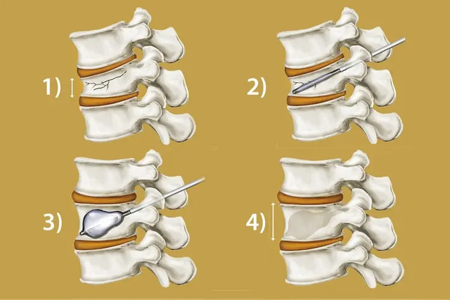 Kyphoplasty — balloon inflated to restore vertebral height before cementing
Kyphoplasty adds an important step: a balloon catheter is inflated inside the fractured vertebra under pressure, restoring vertebral body height before cement injection. The balloon is then deflated and removed, and bone cement is injected into the cavity at low pressure. Advantages: Vertebral height restoration reduces kyphotic deformity, improves posture and respiratory function. Lower cement leakage risk vs. vertebroplasty. Preferred when significant height loss or angular deformity is present.
Who Needs ItIndications
Osteoporotic vertebral compression fractures: Most common indication — post-menopausal women and elderly men. Pathological fractures: From metastatic cancer, multiple myeloma, or haemangiomas. Traumatic fractures: Selected acute compression fractures in patients unsuitable for conventional stabilisation. Most effective for acute/sub-acute fractures within 6 weeks; selected chronic fractures with bone marrow oedema on MRI also respond well.
RecoveryWhat to Expect
Procedure takes 30–60 minutes under local anaesthesia and sedation. Patients sit up within 2 hours, walk the same day, and are typically discharged same day or next morning. Significant pain relief in most patients within 24–48 hours. Return to normal light activity within days. Underlying osteoporosis managed concurrently to prevent future fractures.
Congenital Spinal SurgeryNeural Tube Defects & Congenital Spinal Conditions
Spinal neural tube defects are among the most common serious congenital abnormalities. Expert early surgical management gives children the best possible neurological and functional outcome.
Expert Neonatal & Paediatric Spinal SurgeryGiving Every Child the Best Chance
Spinal neural tube defects require surgical repair that is often urgent — within the first 24–48 hours of life for open defects. Our centre manages the full spectrum under IONM monitoring, in close collaboration with neonatology and paediatric teams. Careful pre-operative counselling of families, clear explanation of expected outcomes, and dedicated post-operative rehabilitation support are integral to our approach.
MyelomeningoceleMyelomeningocele (Open Spina Bifida)
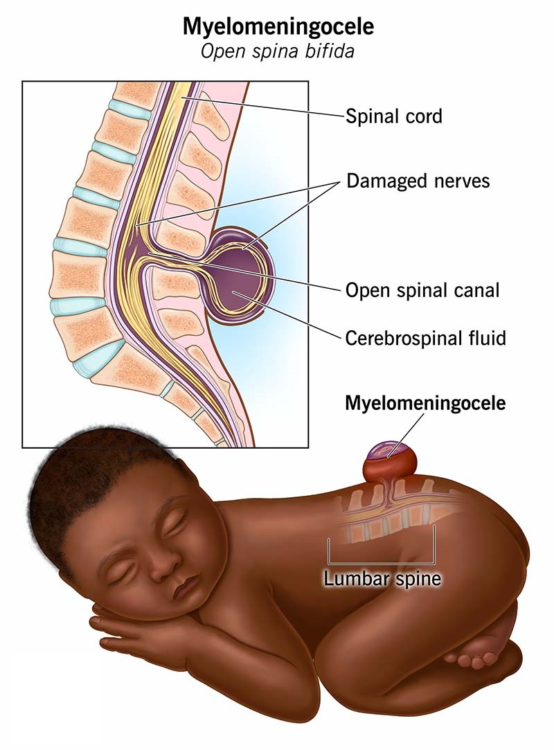 Myelomeningocele — open neural tube defect requiring emergency closure
The most severe form of spina bifida — the spinal cord and nerve roots are exposed on the back through an open defect, covered only by a thin membrane or open to air. This requires emergency surgical closure within 24–48 hours of birth to prevent infection and preserve remaining neurological function. The procedure: The neural placode (exposed spinal cord tissue) is carefully identified, freed from surrounding skin and fascia, and rolled into a tube to reconstruct the neural tube. The dura is closed over it, followed by layered fascial and skin closure. Post-operative monitoring in the NICU is mandatory. Associated conditions: Virtually all myelomeningocele patients have an associated Chiari II malformation and develop hydrocephalus requiring VP shunt or ETV. Bladder, bowel, and lower limb function are assessed and managed by the multidisciplinary team.
MeningoceleMeningocele
 Meningocele — CSF-filled sac with intact overlying skin
A meningocele is a sac of meninges and CSF protruding through a vertebral defect — without neural tissue within the sac. The spinal cord itself is in its normal position. Presentation: A fluctuant, skin-covered or skin-deficient sac on the back, most commonly in the lumbar or sacral region. Most patients with simple meningocele have normal or near-normal neurological function. Surgery: The sac is excised, the neck of the sac is ligated and oversewn with watertight dural closure, and the skin is closed in layers. Because the spinal cord is not involved, outcomes are generally excellent with good neurological preservation.
Tethered CordTethered Spinal Cord Syndrome
 Tethered cord — low-lying conus on MRI
Tethered cord syndrome occurs when the spinal cord is abnormally fixed (tethered) to surrounding structures — most commonly by a thickened filum terminale, lipoma, dermoid cyst, diastematomyelia, or scar tissue from previous neural tube defect repair. As the child grows, the tethered cord is stretched, causing progressive neurological deterioration. Symptoms: Back and leg pain, progressive weakness of legs, bladder and bowel dysfunction, foot deformity, scoliosis. Surgery (cord untethering): Performed under continuous IONM (MEPs, SSEPs, and triggered EMG of bladder and sphincter muscles). The tethering structure is identified through a small laminectomy and carefully divided or excised under microscopic vision — releasing the cord and allowing it to rise to its normal position. Results are often excellent — halting or reversing neurological deterioration. Hair-sparing principles are adapted here: no unnecessary shaving of posterior midline skin hair.
LipomyelomeningoceleLipomyelomeningocele & Spinal Lipoma
 Lipomyelomeningocele — fatty mass attached to spinal cord
A lipomyelomeningocele is a form of closed spinal dysraphism where a fatty (lipomatous) mass is attached to the spinal cord and extends through a bony defect into a subcutaneous mass visible on the back. Unlike open myelomeningocele, the skin is intact. Surgery involves excision of the lipoma as completely as safely possible and untethering of the spinal cord, performed under IONM. The goal is to prevent future neurological deterioration as the lipoma grows and the cord stretches with the child's growth.
DiastematomyeliaDiastematomyelia (Split Cord Malformation)
 Diastematomyelia — split cord malformation on MRI
In diastematomyelia, the spinal cord is split into two hemicords by a bony, cartilaginous, or fibrous spur arising from the vertebral body. The spur tethers the cord, causing progressive neurological deterioration. Surgery involves identifying and excising the spur through a posterior approach, freeing both hemicords and allowing them to reunite. IONM is used throughout to protect both hemicords during the procedure.
Dermal SinusCongenital Dermal Sinus
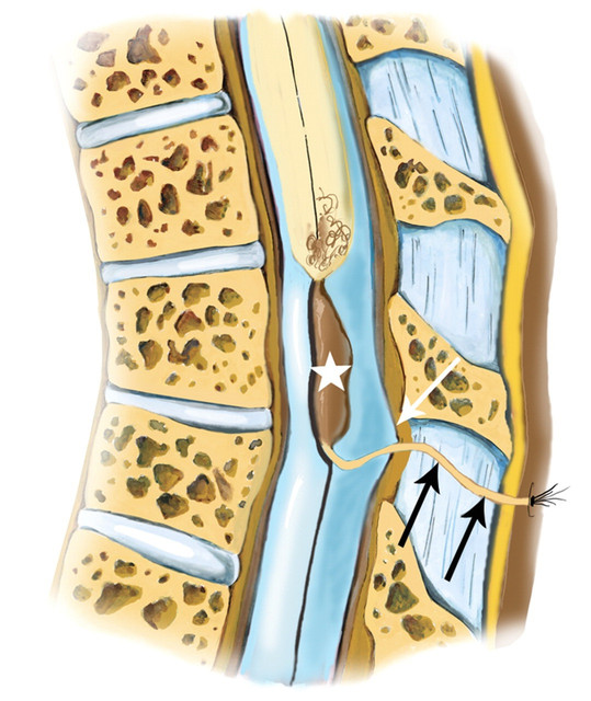 Dermal sinus tract — route for meningitis if untreated
A congenital dermal sinus is a tract of skin-lined tissue extending from the skin surface inward toward the spinal canal — often associated with a dermoid or epidermoid cyst at its inner end. It is a potential route for bacterial meningitis and requires excision of the complete tract before infection occurs. Surgery involves following the tract through a laminectomy into the spinal canal, excising it entirely along with any associated intradural cyst, under continuous IONM.
Sacrococcygeal TeratomaSacrococcygeal Teratoma
 Sacrococcygeal teratoma — most common neonatal tumour
A germ cell tumour arising at the sacrococcygeal region — the most common neonatal tumour. Mature teratomas are benign but require complete surgical excision including the coccyx to prevent recurrence. Immature and malignant teratomas require surgery plus adjuvant chemotherapy. Bowel and bladder function monitoring is integral to post-operative care.
Spinal InfectionsSpinal Infections & Tuberculosis
Spinal tuberculosis (Pott’s spine) is the most common form of skeletal TB and a leading cause of spinal deformity and paralysis in India. With early diagnosis, modern medical therapy, and minimally invasive surgery when needed, the vast majority of patients recover fully — even those who present with weakness.
Cure Is the GoalPreserving the Cord, Correcting Deformity, Eradicating Infection
Most spinal infections are controlled with targeted antibiotic or anti-tubercular therapy and bracing. Surgery is reserved for clear indications — neurological deficit, spinal instability, progressive deformity, large abscesses, or failure of medical treatment. When required, modern techniques allow thorough debridement, decompression of the spinal cord, drainage of abscesses, and stabilisation in a single stage — restoring alignment and protecting function while the infection is cured.
Pott’s SpineSpinal Tuberculosis (Pott’s Spine)
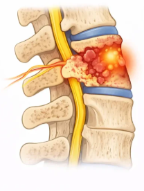 Pott’s spine — vertebral destruction and angular kyphosis
TB most often affects the thoracolumbar spine, destroying the vertebral bodies and discs, forming a “cold abscess,” and producing a sharp angular kyphotic deformity. Untreated, it can compress the spinal cord and cause paralysis (Pott’s paraplegia). Treatment: a full course of anti-tubercular therapy is the foundation. Surgery — debridement, cord decompression, and instrumented fusion (anterior, posterior, or combined) — is added for neurological deficit, instability, or significant deformity.
Epidural AbscessSpinal Epidural Abscess
 Epidural abscess compressing the spinal cord
A collection of pus within the spinal canal pressing on the cord or nerve roots — a true emergency that can cause rapid, permanent paralysis. Presents with severe back pain, fever, and progressive weakness. Treatment: urgent surgical decompression and drainage (often through a minimally invasive approach) to relieve pressure, combined with prolonged targeted antibiotics. The speed of surgery directly determines whether neurological function is preserved.
DiscitisPyogenic Discitis & Vertebral Osteomyelitis
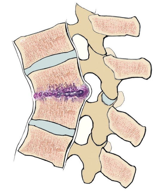 Spondylodiscitis — disc and vertebral infection on MRI
Bacterial infection of the intervertebral disc and adjacent vertebrae, causing intense localised back pain, fever, and stiffness. Most cases are diagnosed by MRI and image-guided biopsy and treated successfully with targeted intravenous antibiotics and bracing. Surgery is indicated for abscess formation, spinal instability, deformity, neurological compromise, or when infection fails to respond to medical therapy — involving debridement and stabilisation.
RecoveryOutcomes & Rehabilitation
With timely treatment, outcomes are excellent — even patients who arrive with weakness frequently regain the ability to walk after decompression and stabilisation. A structured programme of nutrition, physiotherapy, brace support, and completion of the full medication course is essential to prevent recurrence and consolidate recovery. Long-term follow-up confirms eradication of infection and stability of the spine.